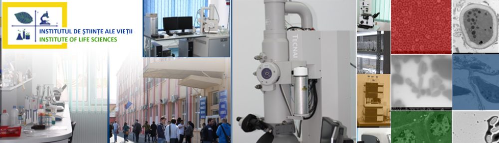
Electron Microscopy Facility
(https://eertis.eu/erlb-2400-001f-1548)
Equipment: Scanning electron microscope – Quanta 250 FEI, Transmission electron microscope – Tecnai 12 Biotwin FEI, Leica EM UC7 Ultramicrotome, AGAR Sputter Coater (gold target) B7340, AGAR Auto carbon coater B7367A
Activities/techniques: transmission (TEM) and scanning electron microscopy (SEM), respectively carrier nanostructure imaging/morphometry analysis for active principles, fungal/dermatophyte species imaging/morphometry analysis, structural and ultrastructural analysis of biological samples from animal and plant tissues, electron tomography and 3D reconstruction of structures, ultrastructures, imaging analysis/cell morphometry/cell lines
Histopathology, Optical, Fluorescence and Confocal Microscopy Facility
(https://eertis.eu/erlb-2400-001u-1556)
Equipment: Olympus BX43 Upright Fluorescence Microscope with Cell Dimension Software, Leica TCS SP8 Confocal Microscope, Mnt SLEE Fully Automatic Cryostat, MicroTec and Leica manual microtomes, Vibrating Blade Microtome Leica VT1000 S, Drying oven SLN 53, histological slide warmer, Motic PA53FS6 Digital Microscope with epifluorescence module, Milestone LOGOS-EVO Advanced Tissue Processor, MYR Paraffin embedding station, Myr – EVA SS-30H Automated slide stainer, Signature Slide Printer, Motic Panthera Cloud Binocular Digital Microscope, Motic Easy Scan Pro 6 Slide scanner, Digital equipment with accessories for sectioning non-calcified bone structures LAM PLAN.
Activities/techniques: histology, histochemistry, immunohistochemistry, immunofluorescence and confocal analysis of animal tissues, frozen and paraffin embedded
Molecular Biology
(https://eertis.eu/erlb-2400-001s-1551)
Equipment: QuantStudio 5 Real-time PCR (Thermo Scientific), PCR T Advanced (Biometra) PCR 2720 Thermal Cycler (Applied Biosystems), Real-Time PCR Rotor-Gene Q (Qiagen), BioDoc-It Imaging System (UVP) imaging documentation system with transilluminator, gel migration system for nucleic acids (3 power sources, 3 horizontal migration tanks), protein gel migration system (PowerPack Basic power supply, a Mini-Protean Tetra System vertical PAGE gel electrophoresis tank, both from Bio-Rad), Percellys 24 Tissue Homogenizer (Bertin Technologies), Tecan Infinite F200 plate reader, Trans-Blot Turbo rapid protein transfer system (Bio-Rad), ChemiDoc MP Imaging system (Bio-Rad), Elisa StatFax 2200 incubator shaker (Awarness Technology), Auxiliary equipment: MS2 Vortex (Ika), pH meter (Hannah), water bath (Julabo), analytical balance (Adam), dry Thermoblock TDB-120 (Kisker Biotech), UV Transiluminator (UVP)
Activities/techniques: extraction of DNA from bacteria, plant, animal and human tissue, animal and human blood samples, extraction of RNA from bacteria, plant, animal and human tissue, animal and human blood samples, standard PCR, Real Time PCR, Single Nucleotide Polymorphism (SNP) and High Resolution Melt (HRM) analysis, cloning a fragment, gene of interest into an expression vector to evaluate the function of that gene, evaluating the expression of a protein by labeling it with specific antibody (Western Blot).
Animal/human cell culture
Equipment: Laminar flow hood, CO2 incubator, Kruss inverted microscope, Motic AE2000 phase contrast digital inverted microscope , Tecan plate reader, centrifuge, water bath, -80°C Ultra Low Lab Freezer, UV lamp.
Activities/techniques: maintaining different cell lines, morphological characterization of cells, determining the cytotoxicity of some phytotherapeutic products by calculating cell viability, etc.
Citogenetics
(https://eertis.eu/erlb-2400-001y-1554)
Equipment: Nikon H600L Microscope (Made in Japan), Meta Class Capture and Karyotyping System (Made in Spain), MiDI 40 CO2 Thermostat – Incubator
Activities/techniques:
Cytogenetic analysis: standard karyotype by GTG banding at 550 band resolution; karyotype at 850 band resolution, for couples with reproductive failures (abortions, stillbirths, male oligo-astheno-teratozoospermia, female sex with primary-secondary amenorrhea, premature menopause, for newborns with particular phenotype, newborns with sexual ambiguity, overweight children, children with mental retardation, autistic manifestations, for leukemias in hemato-oncology.
DNA fragmentation test for male idiopathic oligo-astheno-teratozoospermia with karyotype and normal Y chromosome deletions.
Ecology
Equipment:
Activities/techniques:
Flowcytometry
Equipment: FC500 flow cytometer from Beckman Coulter
Activities/techniques: analysis of cell viability, cell death, cell proliferation and cell cycle, diagnosis of onco-hematological diseases
High performance liquid chromatography
Equipment: Ultimate 3000 HPLC (Dionex)
Activities/techniques: separation, identification and quantification of compounds of interest; analysis of plant extracts (crude or powdered extracts) for determination of flavonoids, amino acids, alkaloids
Animal Research Facility
(https://eertis.eu/erlb-2400-001d-1543)
The Animal Facility is an authorized research infrastructure for breeding and conducting of experiments on laboratory animals authorized by the National Sanitary Veterinary and Food Safety Authority (ANSVSA authorization 1475/15.04.2021).
Equipment: IVC (Individually Ventilated Cages) housing systems for mice and rats; two Animal Cage Changing Stations provided with laminar flow and HEPA filters; Functional Explorations Lab equipped with BS-120 Chemistry Analyzer and Urit-2900 Vet Plus Auto Hematology Analyzer; operating and treatment room; InVivo Xtreme Preclinical Optical/X-ray Imaging System for preclinical studies that allows the following imaging modalities: luminescence, fluorescence, radioisotopic and radiographic imaging and the analysis of bone densitometry, In vivo Fluorescence Imaging System FOBI 2 Blue Green Red and NIR Channels – Cell Gentek – FM21-BGRN, Laser Speckle Imaging System – RWD Life Science – RFLSI III, Nanoliter Injection Pump RWD Life Science – R-480
Activities/techniques: The maximum housing and breeding capacity of the Animal Research Facility is: a) mice –1050 animals b) rats – 280 animals c) hamsters – 250 animals; in the Functional Exploration Laboratory different tests are performed for health monitoring of the animals (hematological and biochemical analysis); in the operation and treatment room are performed: general anesthesia by inhalation and by injection, oral administration (gavage), intraperitoneal, intramuscular, intravenous and intraarterial administrations, organ perfusions, organ harvesting, surgical interventions (creation of bone defects and replacing it with a scaffold), postoperative clinical monitoring. In vivo imaging allows to perform in vivo pharmacodynamic studies, testing and validation of samples and biomarkers; in vitro and in vivo monitorization of cell migration; simultaneous image recording (optical and radioisotopic) with X-ray images.
Plant Biotehnology
(https://eertis.eu/erlb-2400-001e-1557)
Equipment: laminar flow hood, autoclave, bidistillator, oven, magnetic stirrers, glassware, lighting sources (LED, phosphorescent materials, colored fluorescent tubes).
Activities/techniques: The research activities are focused on the application of in vitro technologies to create different plants with valuable characteristics, which are hard to obtain by traditional methods.
An important direction of investigation involves the obtaining and multiplication of virus-free plants by callusogenesis and the use of the embryonic meristematic tissues.
Based on the research carried out during many years of study, different protocols were developed to obtain plant species with therapeutic and ornamental character (Sedum telephium ssp. maximum L., Occimum basilicum var. Aromat de Buzău, Lycium barbarum) as well as healthy planting material for fruit trees and shrubs. Solidification agents more efficient than agar are used to obtain the culture medium, especially from an economic point of view. Efficient lighting equipment, such as LEDs of different wavelengths and phosphorescent materials are used to illuminate the in vitro cultures.
The “Pavel Covaci” University Botanical Garden of the “Vasile Goldiş” Western University of Arad was founded in 1968 as Macea Dendrological Park (since 1994 – Botanical Garden, since 2010 – “Pavel Covaci” University Botanical Garden). It has an area of 21,5 ha and has over 3.800 taxa in its collection. The “Pavel Covaci” University Botanical Garden represents an important research base of the ”Aurel Ardelean” Institute of Life Sciences. The Botanical Museum is housed in an old hunting lodge near the Castle, which also includes the Seed Collection, the Herbarium and two research laboratories in the field of plant biology.
The “Pavel Covaci” University Botanical Garden in Macea is a founding member of the Association of the Botanical Gardens in Romania (AGBR) headquartered in Bucharest and a member of the Botanic Gardens Conservation International (BGCI) headquartered in Richmond (England). The garden is an institution that has attributions in the process of learning, research and public education in order to conserve the biodiversity. It has many scientific connections and has permanent exchanges of biological material with over 225 botanical gardens from Romania and abroad. (https://eertis.eu/erlb-2400-001t-1669)